
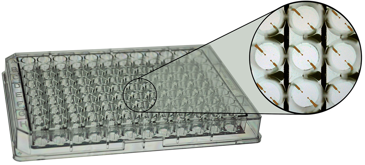
ECIS Cultureware consists of sterile disposable electrode arrays containing gold film electrodes delineated with an insulating film. The well top assembly is made of polystyrene. The gold layer is thin enough to allow microscopic observation of the cells using a standard inverted tissue culture microscope. The ECIS electrode array is placed in an array holder located in the incubator.
For help in chossing an ECIS array we offer a technical note for download - TN01 Array Choices
| Array | Electrodes per Well | Std. Filter Area (cm2) | Maximum Number of Cells Measured | Well Volume (µL) |
|---|---|---|---|---|
| 24WTEER PET | 1 | 0.33 | 30,000-40,000 | 1000, 200 (transfilter) |
| Array | Electrodes per Well | Electrode Area (mm2) | Maximum Number of Cells Measured | Well Volume (µL) |
|---|---|---|---|---|
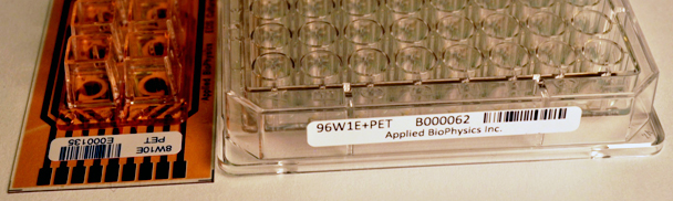
| 96W1E+ PET | 2 | 0.256 | 100-200 | 300 |
| 96W10idf PET | idf | 2.09 | 2000-4000 | 300 |
| 96W20idf PET | idf | 3.985 | 4000-8000 | 300 |
| Array | Electrodes per Well | Electrode Area (mm2) | Maximum Number of Cells Measured | Well Volume (µL) |
|---|---|---|---|---|
| 8W1E PET or PC | 1 | 0.049 | 50-100 | 600 |
| 8W10E PET or PC | 10 | 0.49 | 500-1000 | 600 |
| 8W10E+ PET or PC | 40 | 1.96 | 2000-4000 | 600 |
| 8W20idf PET | idf | 3.985 | 4000-8000 | 600 |
| Array | Electrodes per Well | Electrode Area (mm2) | Maximum Number of Cells Measured | Well Volume (µL) |
|---|---|---|---|---|
| 8W2x1E PET or PC | 2x1 | 2 x 0.049 | 50-100 | 600 |
| 8W1CXE PET or PC | 1 | 0.049 | 50-100 | 600 |
| 8W2LE PET or PC | 2 | 0.20 | 200-400 | 600 |
| Array | Electrodes per Well | Electrode Area (mm2) | Maximum Number of Cells Measured | Well Volume (µL) | Channel Height x Width (mm) |
|---|---|---|---|---|---|
| 1F8x1E PC | 8x1 (1 channel) | 0.049 | 50-100 | 90/60 | 0.36 x 5 |
| 1F8x10E PC | 8x10 (1 channel) | 0.49 | 500-1000 | 90/60 | 0.36 x 5 |
| 6F1E PC | 1 (6 channels) | 0.049 | 50-100 | 45/60 | 0.66 x 5 |
| 6F10E PC | 10 (6 channels) | 0.49 | 500-1000 | 45/60 | 0.66 x 5 |
Small electrodes (1E, 10E, 10E+ type arrays) and their layout within the wells ensure that all current passes through the cell monolayer. This allows the ability to analyze data with the ECIS modeling software to determine barrier function, cell membrane capacitance as well as the spacing between the cell basal membrane and electrode.
Keeping the total surface area of the electrodes small also allows for a relatively low AC current to generate the large electric field necessary to either electroporate or kill the cells in migration experiments. Small electrodes also provide the ability to monitor the uncorrelated nano-scale morphological changes of individual or small populations of cells (<100), while larger or multiple electrodes provide the averaged morphological response of many cells (1000+).
Some experimental protocols, such as cell proliferation, require sparse inoculations leading to a variance of cell density at the bottom of the well. Large electrodes (CP Array) or a large collection of small electrodes (10E+ Array) increases the sampling size resulting in less variability.
Applied Biophysics offers arrays optimized for observing cells under a variety of conditions such as high shear stress and hypoxia/hyperoxia. Additionally, specialized arrays for correlated microscopy and ECIS are available.
PET: Polyethylene terephthalate, standard thickness 0.25mm
PC: Clear polycarbonate substrate, standard thickness 0.51mm
High numerical aperture (HNA) series with a thickness of 0.13mm is available - contact factory to order
All arrays include a bar code (code 128) label and serial number. The ECIS software embeds the type of array and serial number information in the ECIS data file. The information can be scanned in via a bar code scanner or keyed in manually. Requires ECIS software V 1.2.135 or later.
We maintain a large supply of arrays for shipment from our facility. When arrays are ordered, a final oxygen plasma etching procedure is used to both clean the gold surfaces and to sterilize the arrays. They are then packed and mailed on the same day via FedEx 2nd day. Credit cards are accepted.
For array ordering and questions please contact Catherine Toniatti at (518) 880-6860. You may also send an email to ctoniatti@biophysics.com or fax your order with contact numbers and all shipping & billing addresses to 518-880-6865.
All arrays can be ordered without upper chambers (NC). Gold films are transparent and the upper chambers can be removed following ECIS measurements to facilitate staining including fluorescence measurements.
Each of the 24 wells in a standard plate configuration contains a gold electrode covering the entire bottom of each well on a transparent PET substrate. This plate is compatible with any commercially available 24 well sized filter. Filters are not included.
We recommend Corning Transwell® Permeable Supports (#3413). Recommended pore size is 0.4um – however, the plate works with any pore size including tortuous path.
Applications Include:
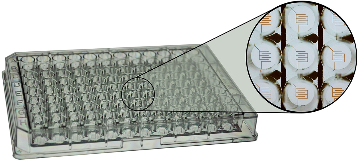
Each of the 96 wells in a standard plate configuration contains two circular 350 μm diameter active electrodes on a transparent PET substrate (measuring from 100-200 cells). As with other 1E arrays, a major use of this array is for the ECIS wound-healing assays where the small electrodes assures the high current pulse will result in complete cell killing.
Only a small population of cells is monitored on the small electrodes resulting in a fluctuating impedance signal due to the random like movement of the cells (micromotion).
Applications Include:
Each of the 96 wells has an Inter-digitated finger configuration. The total electrode area is 2.09mm2 which measures a maximum of 2000-4000 cells.
Applications Include:
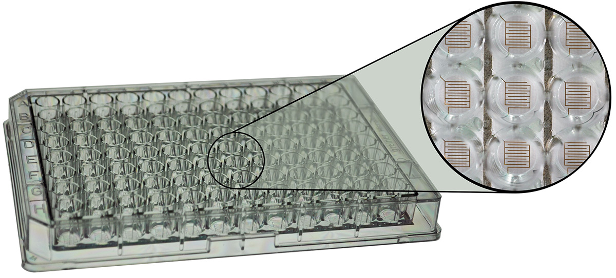
Each of the 96 wells has an Inter-digitated finger configuration. The total electrode area is 3.985mm2 which measures a maximum of 4000-8000 cells.
Applications Include:
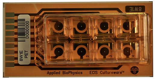
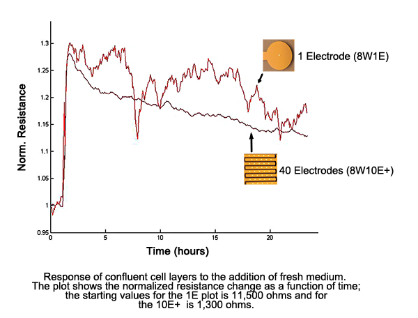
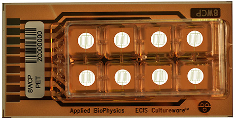
Each of the 8 wells contains a single circular 250μm diameter active electrode. Each well has a substrate area of 0.8 cm2 and a maximum volume of 600μL. On average, with a confluent cell layer, approximately 50 to 100 cells will be measured by the electrode, but even a single cell can be observed.
Applications Include:
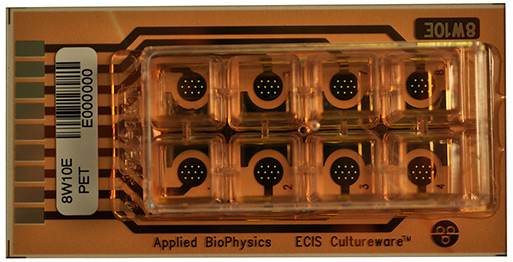
Each of the 8 wells contains ten circular 250 μm diameter active electrodes connected in parallel on a common gold pad. Each well has a substrate area of 0.8 cm2 and a maximum volume of 600 μL. On average, with a confluent cell layer, approximately 500 to 1000 cells will be measured by the electrodes.
Applications Include:
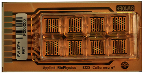
Each of the 8 wells has two sets of 20 circular 250 μm diameter active electrodes located on inter-digitated fingers to provide measurements of cells upon a total of 40 electrodes. Each well has a substrate area of 0.8 cm2 and a maximum volume of 600 μL. On average, with a confluent layer, approximately 2000 to 4000 cells will be measured by the electrodes.
The 10E+ arrays are designed to monitor larger numbers of cells, sampling over the entire bottom of the well. Because of the relatively high number of cells, impedance fluctuations due to micromotion are smoothed out and do not obscure subtle changes in impedance due to the experimental conditions.
Applications Include:
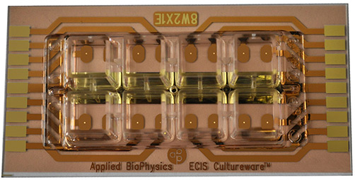
Each of the 8 wells has a total electrode area of 3.985mm2 located on inter-digitated fingers to provide measurements of cells. Each well has a substrate area of 0.8cm2 and a maximum volume of 600 μL. On average, with a confluent layer, approximately 4000 to 8000 cells will be measured by the electrodes.
The 8W20idf PET arrays are designed to monitor larger numbers of cells, sampling over the entire bottom of the well. Because of the relatively high number of cells, impedance fluctuations due to micromotion are smoothed out and do not obscure subtle changes in impedance due to the experimental conditions.
Applications Include:
This array is also called the Medusa array. Each well in this array has two independent single 250 µm diameter active electrodes. The Medusa array is useful for duplicating readings in the same well or to wound/electroporate one electrode while leaving the other as a control within the same well.
When connected to the array holder only the upper four wells are measured. To use the other four wells, the array is turned around and the contact pads at the other end are connected.
Applications Include:
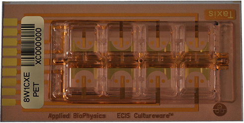
This array is used to monitor the movement of cells in response to chemical gradients and is the array used in chemotaxis measurements first described by Hadjout, N. et al. (2001) Biotechniques 31 (5) 1130. The measuring electrode in this array is a thin gold line between two registry marks.*
Each well has a substrate area of 0.8 cm2 and a maximum volume of 600 μL. On average, with a confluent layer, approximately 50 to 100 cells will be monitored by the electrode.
*The gold line has the same total area as a 250 μm single circular electrode.
Applications Include:
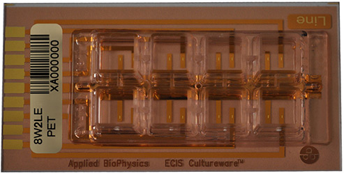
Each of the 8 wells contains a single linear electrode with dimensions of 667µm x 150µm and a measurement value equal to that of our standard 250µm circular electrodes. Each well has a substrate area of 0.8cm2 and a maximum volume of 600μL. On average, with a confluent cell layer, approximately 200 to 400 cells will be measured by the electrode, but even a single cell can be observed.
Applications Include:
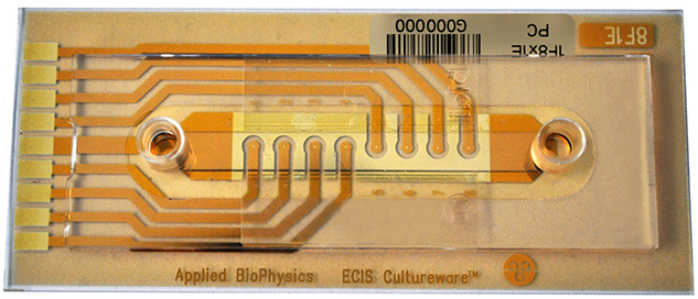
This is a specialized Flow array having 8 active 250 μm diameter electrodes (each measuring from 50-100 cells) located in the central region at the base of a flow channel measuring 50mm in length 5mm in width and available in 0.36mm in height with a total channel volume of 90 μL.
Our flow arrays are designed for ECIS measurements of cells under perfused conditions or to mimic the shear stress endothelial cells experience in vivo or under flow mimicking the shear stress endothelial cells experience in vivo.
Applications Include:
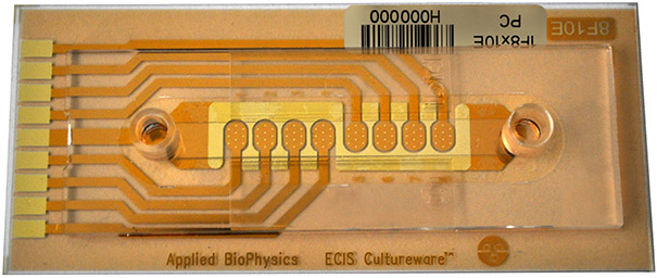
This is a specialized Flow array having 8 sets of 10 active 250 μm diameter electrodes (each measuring from 500-1000 cells) located in the central region at the base of a flow channel measuring 50mm in length 5mm in width and available in 0.36 mm in height with a total channel volume of 90 μL.
Our flow arrays are designed for ECIS measurements of cells under perfused conditions or to mimic the shear stress endothelial cells experience in vivo or under flow mimicking the shear stress endothelial cells experience in vivo.
Applications Include:
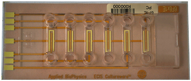
This flow array allows 6 independent flow assays to be run simultaneously. The channels are 0.66mm in height and 5mm wide with 1 active 250 μm diameter electrode (measuring from 50-100 cells) per channel.
Each channel has a 45 μL volume with 60 μL reservoirs.
Applications Include:
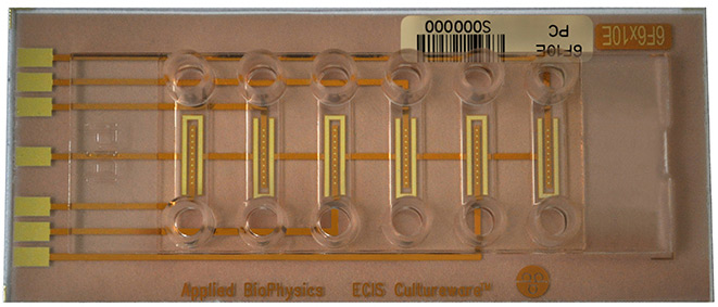
This flow array allows 6 independent flow assays to be run simultaneously. The channels are 0.66mm in height and 5mm wide with 10 active 250 μm diameter electrodes (each measuring from 500-1000 cells) per channel.
Each channel has a 45 μL volume with 60 μL reservoirs.
Applications Include:
© 2026 Applied BioPhysics, Inc.
185 Jordan
Road Troy, NY 12180 / Phone: 518-880-6860 / Toll Free: 866-301-ECIS (3247) / Fax: 518-880-6865